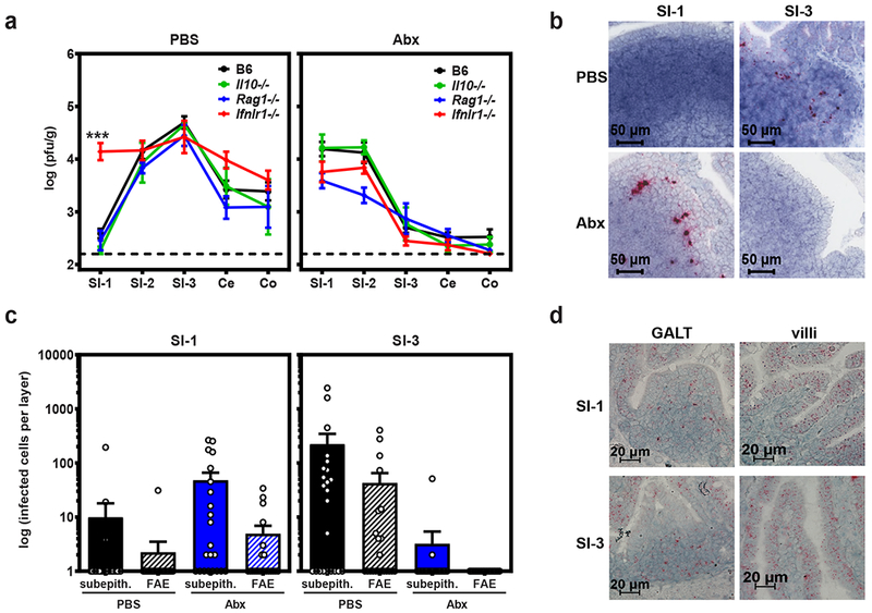
Figure 2. Bacteria-dependent type III IFN signaling in the proximal gut protects intestinal immune cells from MNV-1 infection.

a) Groups of wild-type B6 mice (n = 18) or mice deficient in IL-10 (n = 4), Rag1 (n = 6 for PBS, n = 7 for Abx), or Ifnlr1 (n = 9 for PBS, n = 11 for Abx) were treated with PBS or Abx and then infected with 107 TCID50 units MNV-1. At 1 dpi, virus titers were determined in the indicated segments of the intestinal tract. P <0.0001 for SI-1 titers in B6 versus Ifnlr1−/− mice; titers in all other intestinal segments in knockout mice were statistically similar to titers in B6 mice in both PBS and Abx groups. Unpaired two-tailed Student’s t-tests were used for statistical purposes. ***P < 0.001. b) Groups of B6 mice were treated with PBS or Abx, infected with mock inoculum (n = 4 per condition) or 107 TCID50 units MNV-1 (n = 6 per condition), and intestinal segments harvested at 1 dpi for the purpose of RNAscope-based ISH. Serial sections were hybridized with a probe specific to the minus-strand MNV-1 antigenome indicative of productive infection. Representative images are shown. c) The relative contribution of subepithelial cell (subepith.) versus follicle-associated epithelium (FAE) infection, as indicated by minus-strand viral RNA signal, was quantified as described in the Methods. d) Serial sections from SI-1 and SI-3 of mock-inoculated PBS-treated mice (n = 5 per condition) were hybridized with a probe specific to the Ifnlr1 mRNA and analyzed by RNAscope-based ISH. Representative images from intestinal villi and areas of GALT are shown for each condition. For panels a and c, error bars indicate the mean of all data points.
