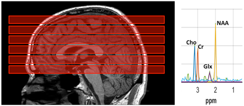
Figure 1.
Sagittal T1-weighted MRI of the brain (left) of a participant with autistic spectrum disorder (ASD) shows prescription of the 6 multiplanar chemical shift imaging (MPCSI; repetition-time [TR]/echo-time [TE]=2800/144 ms) slabs (red blocks), each parallel to the anterior commissure–posterior commissure (AC-PC) plane. Each slab was 10 mm thick with 2-mm interslice gap. One slab passed through the AC-PC, one was just inferior to it, and 4 slices were superior to the AC-PC. In-plane, nominal MPCSI voxel size was 10×10 mm2. Lipid-suppression was achieved by 8 extracranial saturation bands (not shown). Typical MPCSI spectrum from an individual 10×10 mm2 voxel (right) plotting radio-frequency signal intensity vs. chemical-shift in parts-per-million (ppm). Well-resolved resonances are seen for N-acetyl-compounds (NAA), glutamate+glutamine (Glx), creatine+phosphocreatine (Cr), and choline-compounds (Cho). Note narrow bandwidth, high signal-to-noise ratio (SNR), and flat baseline uncontaminated by extracranial lipids, macromolecules, or unsuppressed water.

