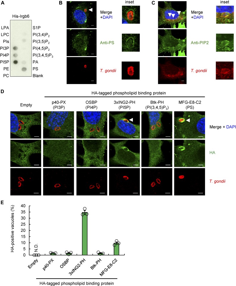
Figure 4. Irgb6 recognizes PI5P and PS at T. gondii PVM.
(A) The representative image from three independent experiments of PIP strips showing the binding of His-tagged recombinant Irgb6 protein to PI3P, PI4P, PI5P, and PS. (B) Confocal microscope images from two independent experiments of the localization of PS (green) with T. gondii (red), and DAPI (blue) in WT MEFs. Scale bars, 5 μm. (C) Confocal microscope images from three independent experiments of the localization of PIs (green) with T. gondii (red), and DAPI (blue) in WT MEFs. Scale bars, 5 μm. (D, E) Confocal microscope images (D) and the graphs (E) from three independent experiments represent the localization of the indicated HA-tagged probes recognizing each specific PIs (green) with T. gondii (red), and DAPI (blue) in WT MEFs. Scale bars, 5 μm. White arrowheads indicate colocalization.

