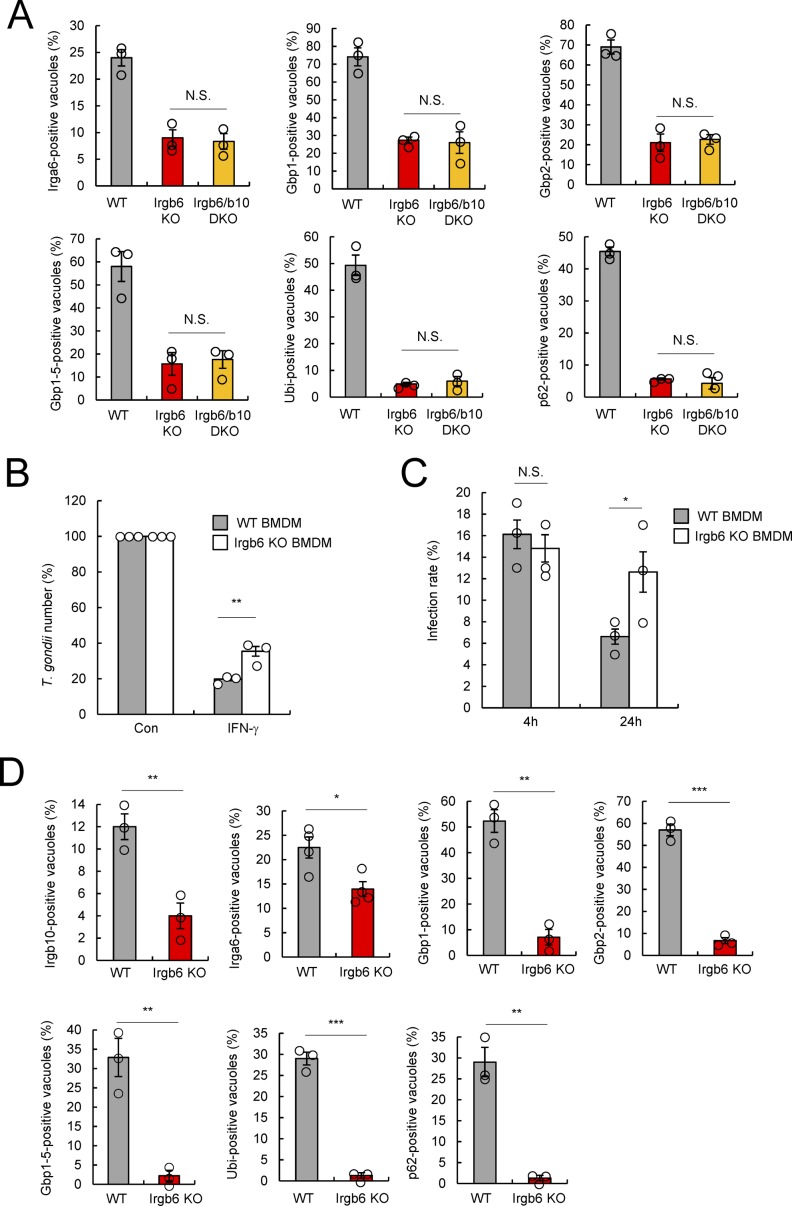
Figure S3. Irgb6 involves in the cell-autonomous immunity of T. gondii in BMDM.
(A) Quantification analysis for Irga6-, Gbp1-, Gbp2-, Gbp1-5-, ubiquitin-, or p62-positive T. gondii vacuoles by confocal microscopy at 4 h postinfection in IFN-γ–treated WT, Irgb6 KO, or Irgb6/b10 DKO MEFs. (B) The parasite survival rate in WT and Irgb6 KO BMDMs by luciferase analysis at 24 h postinfection in the presence or absence of IFN-γ stimulation. (C) Quantification analysis for the infection rate of parasites in WT and Irgb6 KO BMDMs at 4 and 24 h postinfection in the presence of IFN-γ stimulation. (D) Quantification analysis for Irgb10-, Irga6-, Gbp1-, Gbp2-, Gbp1-5-, ubiquitin-, or p62-positive T. gondii vacuoles by confocal microscopy at 4 h postinfection in IFN-γ–treated WT and Irgb6 KO BMDMs. (A, B, C, D) All graphs are means ± SEM from three independent experiments. *P < 0.05, **P < 0.01, and ***P < 0.001 from the two-tailed t test. NS, not significant.

