
Figure 6.
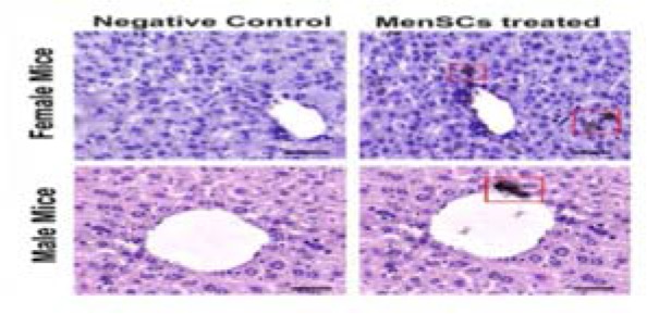
Immunohistochemical staining of mitochondrial protein (red box) in liver samples retrieved from male and female MenSCs-treated groups after 7 days. Liver section from normal mice served as negative control. Scale bar: 100 μm.
Official websites use .gov
A
.gov website belongs to an official
government organization in the United States.
Secure .gov websites use HTTPS
A lock (
) or https:// means you've safely
connected to the .gov website. Share sensitive
information only on official, secure websites.
Immunohistochemical staining of mitochondrial protein (red box) in liver samples retrieved from male and female MenSCs-treated groups after 7 days. Liver section from normal mice served as negative control. Scale bar: 100 μm.