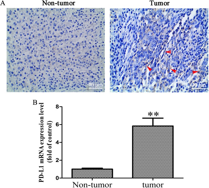
Fig. 1.
Increased PD-L1 expression level in OS tissues. a Immunohistochemical assay was performed to detect the expression of PD-L1 in OS and OS-adjacent tissues (× 400 magnification), the red arrow represents positive PD-L1 staining. b RT-qPCR was performed to detected the mRNA level of PD-L1 in OS and OS-adjacent tissues (n = 6). The results were presented as the mean ± standard deviation. **P < 0.01. PD-L1, programmed death-ligand 1; OS, osteosarcoma; RT-qPCR, reverse transcription-quantitative polymerase chain reaction

