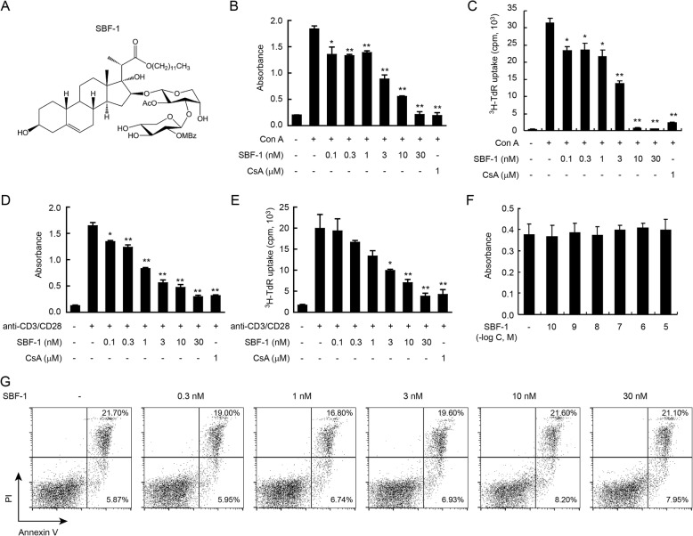
Fig. 1.
SBF-1 markedly inhibited the proliferation of Con A-activated T cells. a The chemical structure of SBF-1. b–e T cells (3 × 10⁵) were isolated from lymph node of BALB/c mice and then incubated with 0.1–30 nM SBF-1 in the presence of 5 μg/ml Con A b–c or 1 μg/ml anti-CD3/CD28 d–e for 72 h at 37 °C. Cell proliferation was measured at 540 nm by MTT uptake assay b, d or [3H]-thymidine uptake assay (c, e). Data are mean ± SEM of three independent experiments. P values were determined by one-way ANOVA with Tukey’s correction. *P < 0.05, **P < 0.01. f–g T cells (5 × 10⁵) were isolated from lymph node of BALB/c mice and then incubated with SBF-1 for 24 h at 37 °C. Cell viability was measured at 540 nm by MTT uptake assay f. Cell apoptosis was analyzed by Annexin V/PI assay staining g. Data are mean ± SEM of three independent experiments

