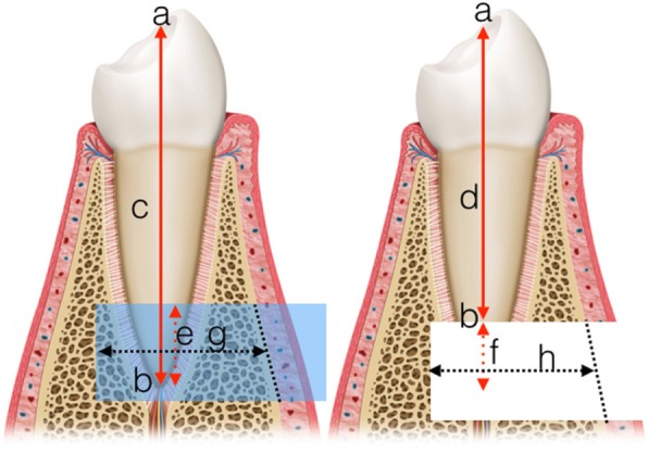
Fig. 4.

Explanation of the 2D measurements. Left: preoperative, Right: postoperative; a: coronal reference point; b: apical reference point (end points of the axis); c: axial length before surgery d: axial length after surgery; e: planned length of removal; f: actual resected length; g: planned depth of osteotomy; h: actual depth of osteotomy (for the measurements, the missing cortical was substituted by a straight line connecting the remaining cortical edges). Calculations: ARE = e-f; ODE = g-h
