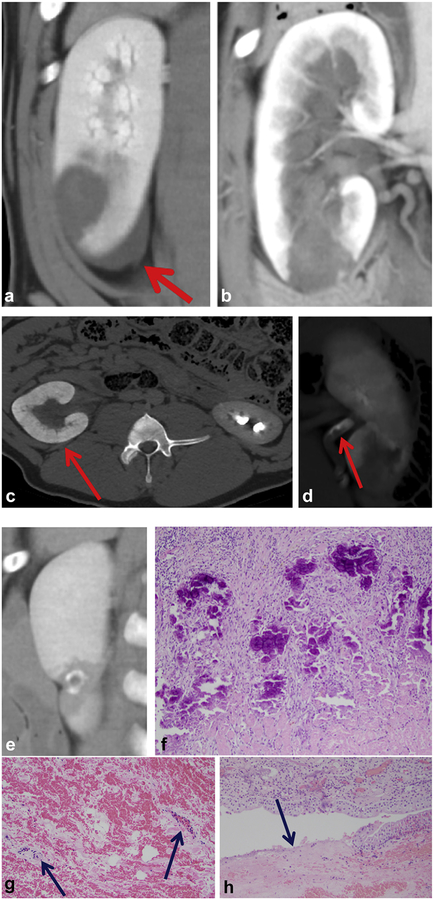
Figure 4.
Various CT images from the study demonstrate common imaging characteristics. (a) Small perirenal fluid collection. (b,c) Transient obstruction of the collecting system. (d) Ureteral debris. (e,f) Calcification of the ablation perimeter on day 28 with corresponding histology (hematoxylin and eosin [H&E], 100X), (g) Preservation of blood vessels with injury within the ablation (H&E, 200X). (h) Occasional disruption of urothelium (H&E, 100X).

