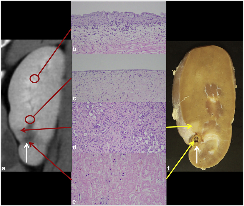
Figure 6.
Chronic treatment. (a) A 4-week post-ablation CT image with small ablation zone (white arrow). (b) Normal urothelium distant from the ablation zone (control, hematoxylin and eosin [H&E], 100X). (c) Re-epithelialized urothelium (H&E, 100X). (d) Atrophy in the transition zone extending laterally (H&E, 100X). (e) Central necrosis in the ablation zone (H&E, 200X). (f) Corresponding gross kidney.

