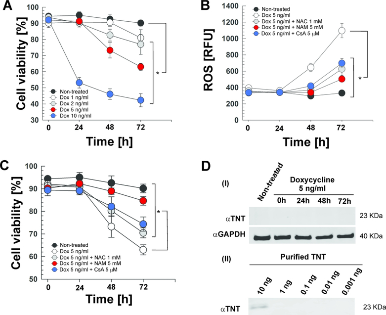
Figure 3. TNT is sufficient to induce lethal oxidative stress in Jurkat-T cells.
Expression of TNT in the Jurkat-T cell line containing an integrated Tet-regulated TNT expression cassette (Jurkat 655-TNT) was induced with different doxycycline (Dox) concentrations at the indicated time points. When indicated, cells were treated with N-acetyl-cysteine (NAC, 1 mM), nicotinamide (NAM, 5 mM) or cyclosporin A (CsA, 5 μM). (A, C) Cell viability was measured by trypan blue staining. (B) ROS levels were measured with the fluorescent probe H2DCFDA. (D) Western blot for (i) TNT (purified specific polyclonal antibody) and GAPDH (loading control) in the whole-cell lysates of Jurkat 655-TNT cells induced with 5 ng/mL doxycycline at the indicated time points, and for (ii) different amounts of purified WT TNT protein to determine the purified specific αTNT polyclonal antibody sensitivity. Asterisks indicate significant differences at 72 h (p-value<0.01, calculated using the One-way ANOVA with Bonferroni’s correction) compared with the indicated conditions. Data are represented as mean ± SEM.

