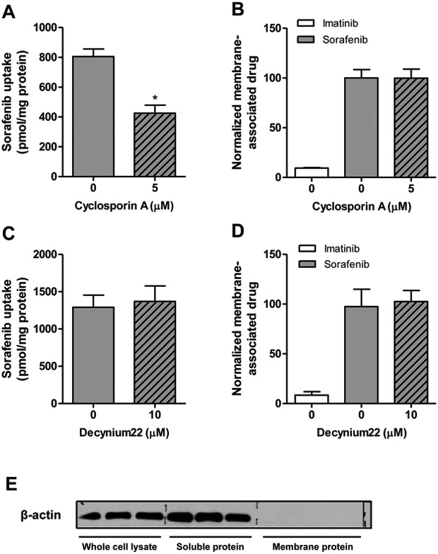
Figure 5.
Membrane binding of sorafenib. (A, B) Sorafenib (0.2 μM) uptake in HeLa cells in the presence and absence of transporter inhibitor cyclosporin A (A) and membrane-associated-drug (B) with or without cycloporin A after 10 min incubations. (C, D) Sorafenib uptake (C) and levels of membrane-associated drug (D) were also evaluated in the presence or absence of decynium22. Imatinib was used as a negative control compound. Data are presented as mean (bars) and SD (error bars) of 3 observations. (E) Expression of β-actin expression in whole cell lysate without membrane isolation, and in soluble protein and membrane protein fractions after membrane isolation as determined by immunoblotting.

