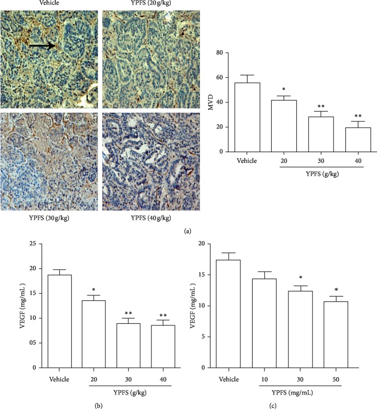
Figure 2.
Effects of YPFS on the angiogenesis of HCC. (a) Tumor tissues from HCC-bearing mice treated with indicated concentration of YPFS (20, 30, and 40 g/kg) were performed by immunohistochemical staining for detecting vascular marker CD34. The pointed tip indicated a positive result and was observed under a 200x microscope. (b) Expression levels of VEGF in tumor tissues were measured by ELISA. (c) Expression levels of VEGF in Hepa1-6 cell were measured by ELISA. Data are presented as the mean ± standard error of the mean. The tumor tissues were observed with a light microscope at ×200 magnification. ∗P < 0.05, ∗∗P < 0.01, compared with the vehicle group.

