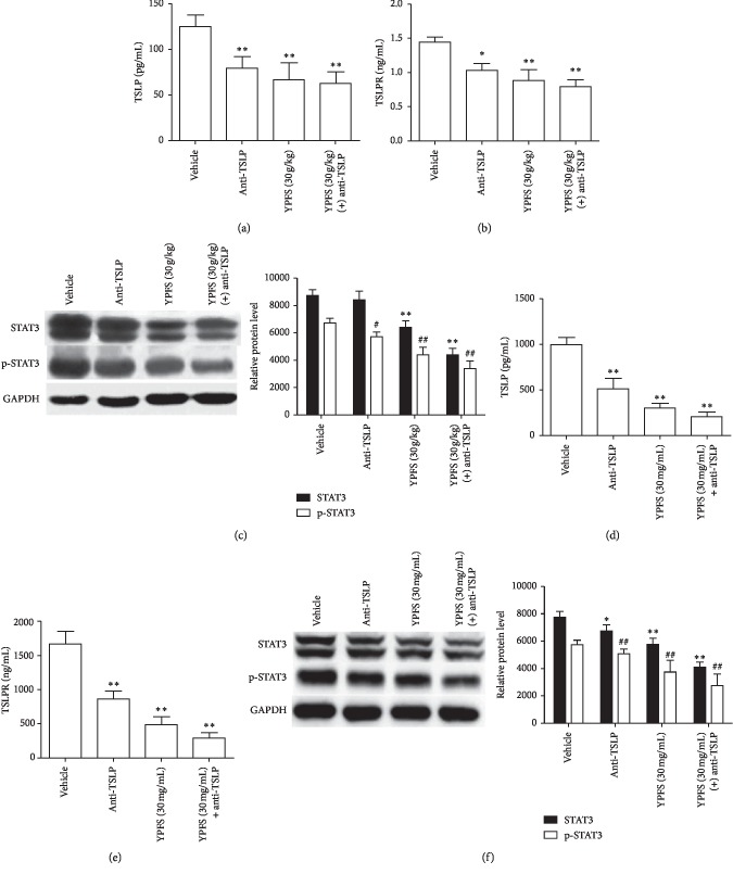
Figure 5.
Anti-TSLP antibody can attenuate the inhibitory effect of YPFS on the signal pathways of TSLP/STAT3 in HCC. (a) Expression levels of TSLP in tumor tissues treated with the anti-TSLP antibody and indicated concentration of YPFS (30 g/kg) mice were measured by ELISA. (b) Expression levels of TSLPR in tumor tissues treated with the anti-TSLP antibody and indicated concentration of YPFS (30 g/kg) mice were measured by ELISA. (c) Western blot assays the expression of STAT3 and p-STAT3 in tumor tissues treated with the anti-TSLP antibody and indicated concentration of YPFS (30 g/kg). (d) ELISA analyzed the expression of TSLP in Hepa1-6 cell of the anti-TSLP antibody and indicated concentration of YPFS (30 mg/mL) treatment. (e) ELISA analyzed the expression of TSLPR in Hepa1-6 cell of the anti-TSLP antibody and indicated concentration of YPFS (30 mg/mL) treatment. (f) The expression of STAT3 and p-STAT3 in Hepa1-6 cell of the anti-TSLP antibody and indicated concentration of YPFS (30 mg/mL) treatment detected by western blot. Quantification of western blot analysis of p-STAT3 and STAT3 levels normalized by the levels of GAPDH. Data are presented as the mean ± standard error of the mean. ∗P < 0.05, ∗∗P < 0.01, #P < 0.05, ##P < 0.01 compared with the vehicle group.

