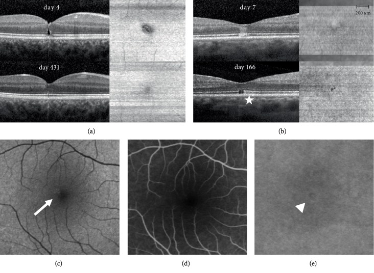
Figure 4.
B‐scan and en face OCT images of patient 2 and 3 (a and b) and multimodal imaging data including autofluorescence imaging (c), fluorescein (d) and indocyanine green (e) angiography of patient 2. (a) OCT B‐scan on day 7 through the foveal center shows a lammelar hole reaching up to the outer nuclear layer; En face OCT shows an EZ alteration boarded by a hyperreflective ring; On day 166 (last follow-up) closure of the lamellar hole and near complete regeneration of the EZ. An inhomogeneous choroidal hypertransmission can be detected until the last follow-up (star). (b) B‐scan OCT on day 4 through the foveal center demonstrates a typical hyperreflective band in the outer retina reaching from the RPE to the outer plexiform layer; there was no distinct hyporeflective zone and no thickening of the outer retinal layers. One month after the incident, B‐scan showed a distinct hyporeflective defect in the ellipsoid zone (EZ), which persisted until last follow-up on day 431. En face OCT on day 4 shows a mild hyperreflective circle, partially surrounded by a scantly hyporeflective zone area; on day 431 a clearly demarcated EZ loss can be observed. (c) Autofluorescence (Heidelberg Spectralis HRA+OCT, excitation at 488nm, barrier filter wavelength of 500nm) showed a hyperautofluorescent zone at first presentation (seven days after the incidence) in the impact region (arrow). (d) Fluorescein angiography (ten minutes after injection) ten days after the incidence did not show any leakage or staining. (e) Indocyanine‐green (ICG) angiography (ten minutes after injection) did show minimal irregular hyper‐ and hypocyanescence in the region of impact (arrowhead).

