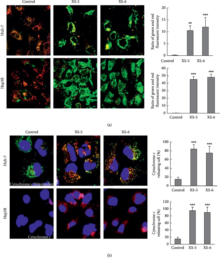
Figure 3.
Effects of XS-5 and XS-6 on mitochondrial apoptosis in HCC cells. (a) When Hep3B and Huh-7 cells were treated with XS-5 and XS-6 (100 μg/ml) for 6 h, the mitochondrial membrane potential was determined by JC-1 staining and analyzed with an Olympus confocal laser scanning microscope. The results (green : red ratio) are expressed as the percentage of cells treated with XS-5 and XS-6. (b) Hep3B and Huh-7 cells were treated with XS-5 and XS-6 (100 μg/ml) for 6 h and were stained with Mitotracker (green) and cytochrome c (red). Localization of cytochrome c in the cytosol was photographed at 400X magnification. Data are expressed as the means ± SD from triplicate experiments (∗∗p < 0.005 and ∗∗∗p < 0.001vs control).

