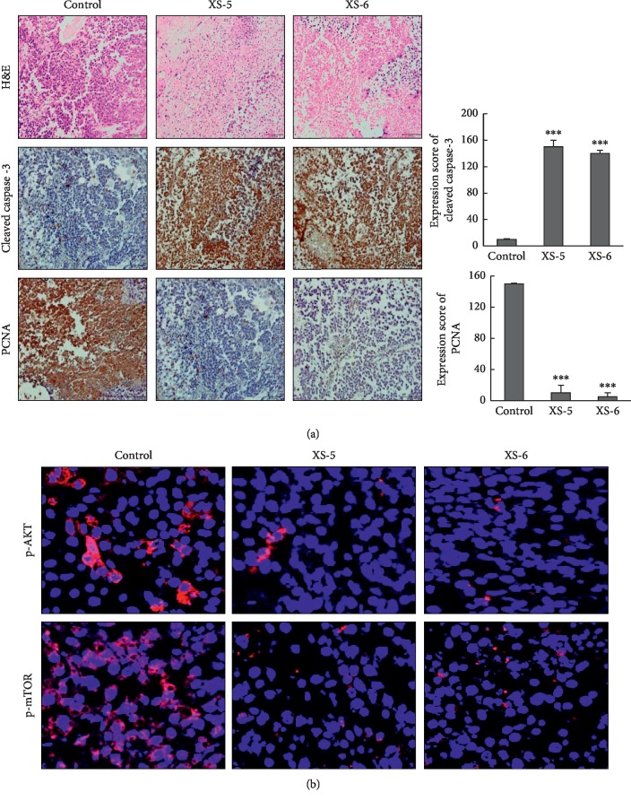
Figure 6.
Effects of XS-5 and XS-6 in HCC ex vivo tumor models. (a) Balb/c nude mice bearing Huh-7 HCC xenograft tumors were cut into small pieces of ∼2 mm, and each piece of tumor was maintained in culture media. Tumor spheroids cultured from xenograft tumor tissues were treated with XS-5 and XS-6 (100 μg/ml) for 5 days. Immunostaining and hematoxylin and eosin staining for PCNA and cleaved caspase-3 were carried out. (b) Tumor spheroids were excised and processed for immunofluorescence for p-AKT and p-mTOR. Images were captured at 400X magnification. Data are represented as the mean ± SD (∗∗∗p < 0.001vs control).

