
Figure 1.
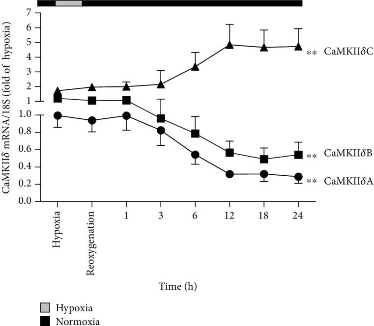
CaMKIIδ variants disordered in cardiomyocytes during H/R injury. After culture in 94% N2, 1% O2, and 5% CO2 for 4 h, the cardiomyocytes were changed into 95% air and 5% CO2. The mRNA levels of CaMKIIδA, CaMKIIδB, and CaMKIIδC of the cardiomyocytes at the start of the hypoxia and different times after reoxygenation were detected by quantitative real-time PCR. 18S was serviced as a housekeeping mRNA. Plots represent the mean ± SEM; n = 6. Statistical significance: ∗∗P < 0.01 compared with “the start of the hypoxia.”