Figure 1.
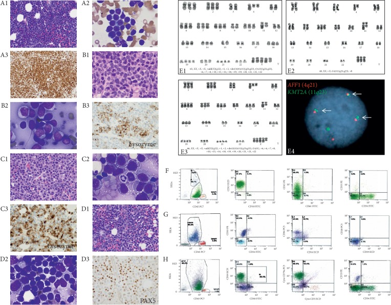
Morphologic, immunohistochemical, flow cytometric, and cytogenetic characteristics of the patient's leukemia. A1–A3 represent bone marrow evaluation at initial diagnosis. Bone marrow biopsy (A1, H&E, 400x) and aspirate (A2, Wright stain 100x, oil) showing numerous small-sized B-lymphoblasts which are strongly positive for PAX5 (A3). B1–B3 represent biopsy of the breast mass (B1, H&E, 400x) and touch imprint (B2, Wright stain, 100x, oil) showing numerous large-sized blasts with monocytic differentiation, which are patchy positive for lysozyme (B3). C1–C3 represent bone marrow biopsy (C1, H&E, 400x) and aspirate (C2, Wright stain, 100x, oil) showing sheets of myeloblasts which are patchy positive for lysozyme (C3). D1–D3 represent bone marrow evaluation six weeks after cessation of blinatumomab. Core biopsy (D1, H&E, 400x) and aspirate (D2, Wright stain, 100x, Oil) show a dimorphic population of blasts: small-sized B-lymphoblasts which are positive for PAX5 (D3) and large-sized myeloblasts which are positive for lysozyme (data not shown here). E1 represents the karyogram of bone marrow specimen at myeloblastic transformation. E2–E4 represent karyograms and AFF1/KMT2A fusion of bone marrow specimen with B/myeloid mixed phenotype acute leukemia. F–H represent flow cytometric features of the leukemic blasts. F represents flow cytometry performed on the bone marrow aspirate at the initial diagnosis showing a large population of B-lymphoblasts (green) in dim CD45 region expressing CD19, CD34 (partial), and CD15 (dim), G represents flow cytometry of the bone marrow aspirate while administration of blinatumomab showing a population of myeloblasts (blue) expressing CD33 and CD64 (dim) and was negative for CD19 and CD34. H represents flow cytometry of the bone marrow aspirate six weeks after cessation of blinatumomab showing two populations: B-lymphoblasts (green) expressing CD19, CD34 (dim), and cytoCD79a, and myeloblasts (blue) expressing CD33, CD64 (dim), and MPO (data not shown here).

