Fig. 3.
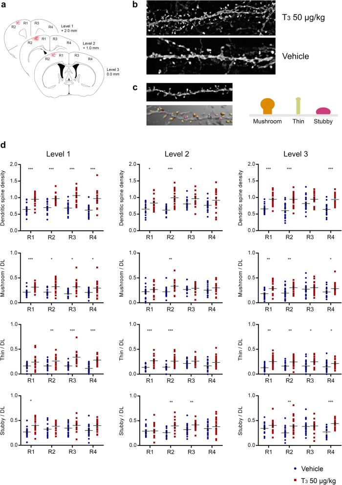
Treatment with T3 50 μg/kg increases dendritic spine density 14 days after photothrombosis (PT). a Dendritic spine analysis 14 days after PT at different distances from bregma correspondent to the rostral pole (level 1), center (level 2) and caudal pole (level 3) of the cortical infarct. The regions analyzed correspond to the ipsilateral (R1) and contralateral (R3) motor cortex; and ipsilateral (R2) and contralateral (R4) somatosensory cortex. b Representative dendritic segments from animals treated with T3 50 μg/kg (n = 4) and Vehicle (Vh; n = 4). c Apical dendritic spines from cortex layers II/III were automatically detected by NeuronStudio software and classified as mushroom, thin or stubby. Three to five dendritic segments were analyzed per animal. d Dendritic spine density (number of total spines / dendritic length) per region and classification of dendritic spines as mushroom, thin or stubby and their density per region, at each level analyzed. Results are displayed as means ± SEM. Statistical analysis was performed with two-tailed unpaired Student t-test, *p < 0.05, **p < 0.01, ***p < 0.001

