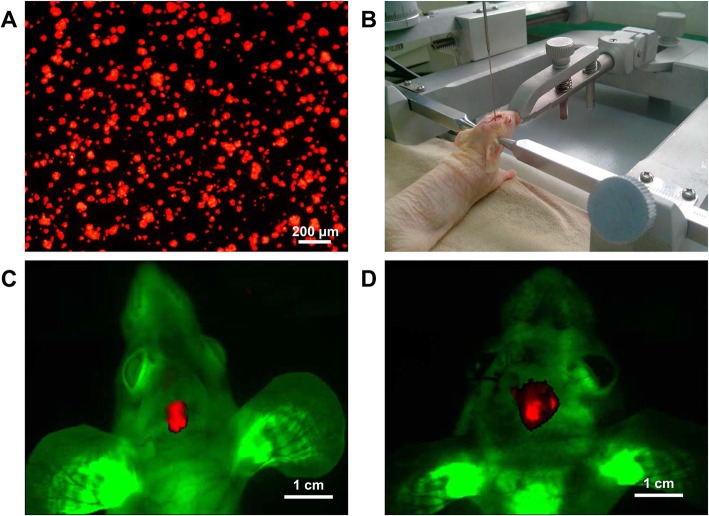
Fig. 1.
Intracranial transplantation of SU3-RFP cells in GFP nude mice. a: SU3 cells were transfected with RFP gene, SU3-RFP cells expressed RFP under fluorescence microscope; b: SU3-RFP cell suspension (1 × 10⁶) was injected into the right caudate nucleus of the mice using a Hamilton syringe with the assistance of a stereotaxic apparatus; c-d: Whole-body images of tumor-bearing mice on day 14 (c) and day 28 (d) after transplantation was obtained using the small animal imaging system (Kodak, USA)

