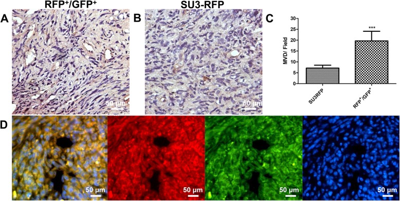
Fig. 9.
Immunohistochemistry to detect CD31 expression in fusion cell xenograft (a) and SU3-RFP xenograft (b); c: Microvessel density comparison between the two groups, MVD-CD31 was 19.67 ± 1.8 vessels in RFP+/GFP + cells and 7.16 ± 0.54 vessels in SU3-RFP cells, respectively; d: Fusion cell xenografts observed under fluorescence microscopy. Scale, A and B 20 μm, D 50 μm. ***p < 0.001

