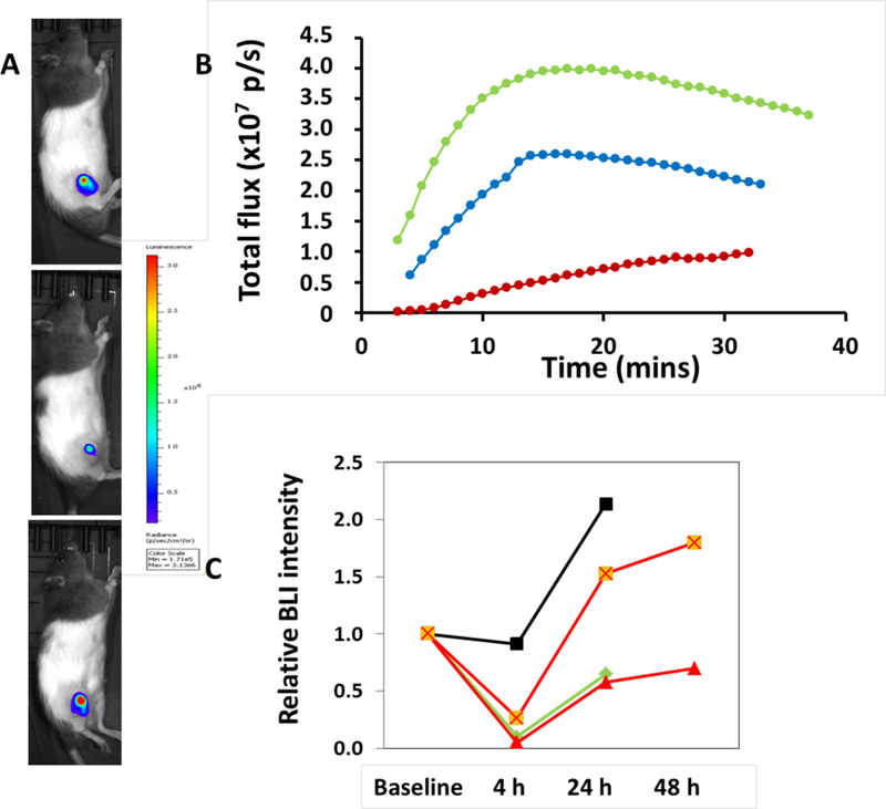
Figure 5.

Relative light emission following administration of VDAs. A) Relative signal intensity is shown about 20 min after administration of D-luciferin subcutaneously in the foreback neck region of a rat with a subcutaneous PC3-DAB2IP-luc prostate tumor xenograft in the thigh. Top) baseline (no prior drug), center) 4 h after 40 mg/kg 69, and bottom) 24 h after 69. B) Corresponding light emission dynamic curve at baseline (blue), 4 h after 69 (red) and 24 h after 69 (green). C) Normalized BLI signal at various times for the rat in Fig 4 receiving 69 sequentially at 10 mg/kg (black), 40 mg /kg (red) and 30 mg/kg CA4P (green), together with the treatment naive rat in Fig 5 A, B receiving 40 mg/kg 69 (orange).
