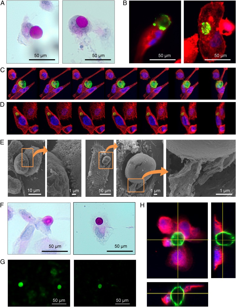
Fig. 5.
CA are phagocytosed by THP-1–derived macrophages. (A) Macrophage in contact with an IgMh-opsonized CA (Left) and an IgMh-opsonized CA phagocytosed by macrophage (Right); PAS staining. (B) Other examples of a macrophage making contact with an IgMh-opsonized CA (Left) and an IgMh-opsonized CA phagocytosed by macrophage (Right). In these cases, macrophages are stained with fluorescent-labeled phalloidin (red) and IgMh-opsonized CA are stained with fluorescent-labeled α-IgMh (green). Nuclei are stained with Hoechst (blue). (C) Sequence of images showing a macrophage phagocytosing an IgMh-opsonized CA from different points of view. The sequence was performed after the 3D reconstruction from images obtained by confocal microscopy. Phalloidin (red), fluorescent labeled α-IgMh (green), and Hoechst (blue). (D) The same procedure was used to obtain a sequence of images showing an IgMh-opsonized CA that has been phagocytosed by a macrophage. (E) SEM images also showed macrophages engulfing IgMh-opsonized CA. Some filopodia were found emerging from macrophages and making contact with the bodies (Insets). (F) Macrophage making contact with a non IgMh-opsonized CA (Left) and a non IgMh-opsonized CA phagocytosed by a macrophage (Right); PAS staining. These results indicate that IgMh opsonization of CA is not a requirement for phagocytosis by THP-1–derived macrophages. (G) CA from CSF become stained with fluorochrome-labeled Con A, which suggests that they contain mannose. (H) CA (stained with Con A, green), encircled by THP-1–derived macrophages immunostained with α-CD206 (mannose receptor, red) and Hoechst (nuclei, blue). Orthogonal views show macrophages attached to the CA. These results suggest that, in the case of THP-1–derived macrophages and with the conditions set up in the present study, CA phagocytosis is produced via the mannose receptor.

