Fig. 1.
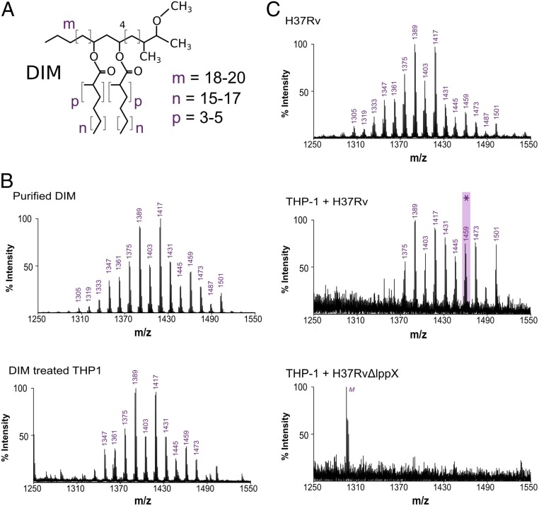
DIMs are transferred from the bacterial envelope to macrophage membranes. (A) Structure of the DIM family of lipids, where m denotes the range of carbon atoms on the phthiocerol moiety, and n and p on the mycocerosate moieties. (B) MALDI-TOF mass spectra of purified DIM and of the membrane fraction of macrophages treated with DIM. (C) MALDI-TOF mass spectra of WT Mtb (HR37v) and of the membrane fraction of macrophages infected by H37Rv or by the H37RvΔlppX mutant. M, low intensity peak corresponding to the detection of the matrix molecule in the DIM region of interest. The asterisk highlights the mass of the DIM molecule chosen for the modeling, with m = 18, n = 17, and p = 4.

