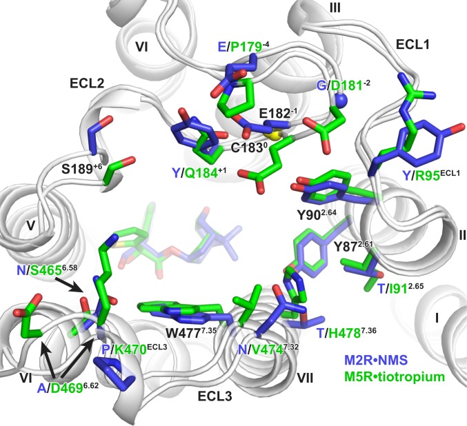
Fig. 3.
Comparison of residues lining the extracellular vestibule of the M2 and M5 mAChR. M2•NMS is shown in dark blue and M5•tiotropium in green. Conserved residues are labeled black, and nonconserved residues have colored labels based on receptor subtype. Residues are numbered based on the M5 mAChR, with residues in ECL2 numbered relative to the conserved cysteine in ECL2, which is shown as a yellow sphere. Sidechains for D4696.62 and K470ECL3 are truncated to the β-carbon in the deposited model due to a lack of sidechain density and are modeled here as the most probable rotamer.

