Fig. 3.
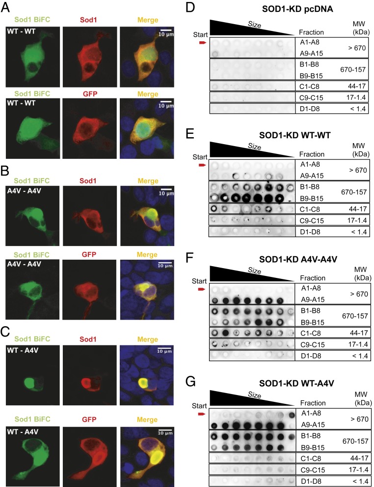
hSod1 WT–A4V heterodimers aggregate into higher molecular weight species. HEK293 Sod1-KD cells were transfected with plasmids encoding the different hSod1 constructs (VN and VC fusions) and imaged using confocal microscopy (A–C). Each panel shows the fluorescence intensities of the BiFC signal counterstained with antibodies against Sod1 or GFP. The anti-GFP antibody used recognizes only the C-terminal fragment of Venus (VC). Sod1-KD cells expressing the homodimer hSod1–WT (A), the homodimer hSod1–A4V (B), and the heterodimer hSod1–WT–A4V (C). SEC followed by dot blot analysis using an anti-hSod1 antibody (D–G). SEC results are representative from at 3 independent experiments. Dot blot from Sod1-KD cells transfected with pcDNA3.1 (D), with WT–Sod1 BiFC constructs (E), with the A4V–Sod1 constructs (F), or with the WT–A4V–Sod1 constructs (G).

