Fig. 3.
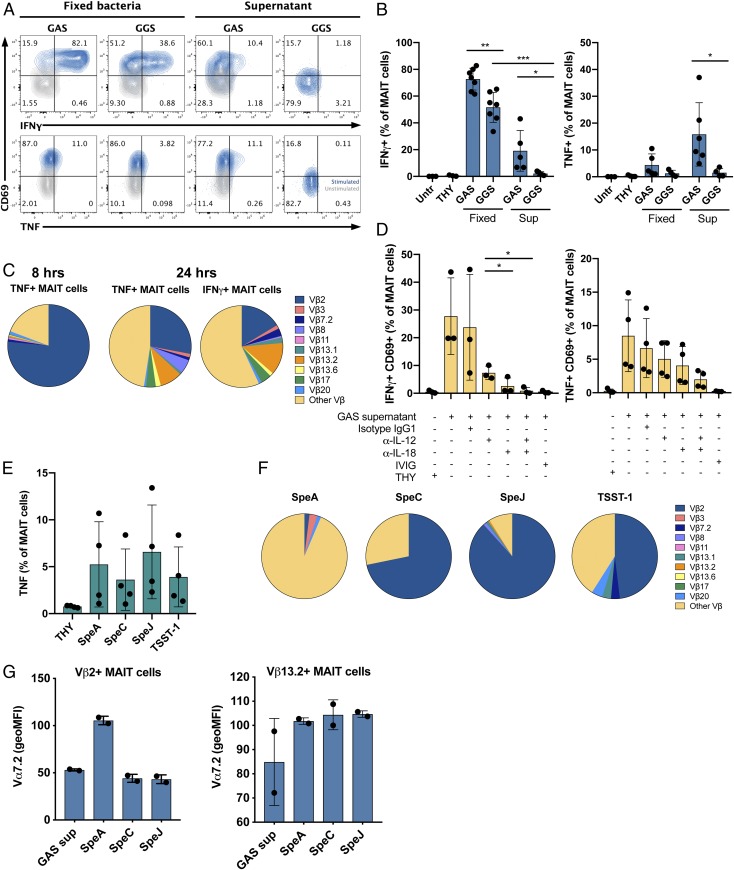
MAIT cells are activated by streptococcal superantigens in a Vβ2-dependent manner. (A and B) Cytokine production by MAIT cells after stimulation of PBMC with fixed bacteria and supernatants of GAS 2006 and GGS 6017 for 24 h assessed by flow cytometry. (A) FACS plot of IFNγ+ or TNF+ MAIT cells from 1 representative donor. (B) Mean frequencies± SD of IFNγ+ or TNF+ MAIT cells of 4 to 7 donors. (C) Stimulation of PBMCs for 8 or 24 h with GAS 2006 supernatant. Vβx expression among cytokine+ MAIT cells was assessed by flow cytometry. (D) Frequencies of IFNγ+ CD69+, or TNF+ CD69+ MAIT cells after stimulation with GAS 2006 supernatant in the presence of anti-IL-12 and/or anti-IL-18 antibody, IgG1 isotype control, or IVIG. Mean ± SD of 3 to 4 donors. (E–G) PBMCs were stimulated with recombinant superantigens and analyzed by flow cytometry. (E) Frequencies of TNF+ MAIT cells after stimulation for 8 h. Mean ± SD of 4 donors. (F) Vβx expression among TNF+ MAIT cells after 8 h of stimulation. Mean ± SD of 2 to 5 donors. (G) Vα7.2 expression (geoMFI normalized to unstimulated control) among Vβ2 or Vβ13.2 MAIT cells after 8 h of stimulation with superantigens of GAS supernatant. Mean ± SD of 2 donors. (B and D) One-way ANOVA was used to detect significant differences between paired samples. ***P < 0.001; **P < 0.01; *P < 0.05.

