Fig. 5.
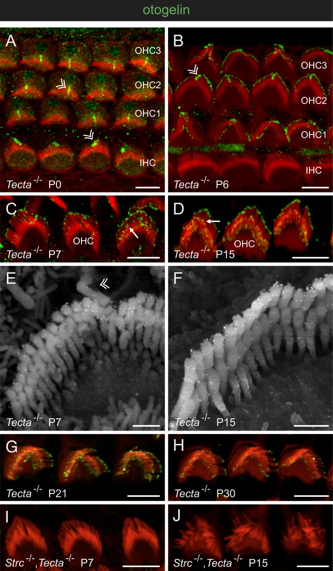
Otogelin in the developing and mature OHCs. Confocal images of whole-mount preparations of the organ of Corti immunostained for otogelin (green) and stained for actin (red) in Tecta−/− mice (A–D, G, and H) and Strc−/−,Tecta−/− double-mutant mice (I and J), and scanning electron micrographs of OHC hair bundles after otogelin immunolabeling in Tecta−/− mice (E and F). In A, B, and E, double arrowheads indicate the transient kinocilium in P0 to P7 IHCs and OHCs (3 rows denoted OHC1, OHC2, and OHC3), which has disappeared on P15. In C and D, arrows indicate the immunostaining in the distal region of middle-sized stereocilia, just above their tip on P7 (C), and in a subapical location corresponding to the position of horizontal top connectors on P15 (D). Immunostained dots are much less abundant on P30 (H) than on P15 (D) and P21 (G), and are not seen in the absence of stereocilin (I and J). (Scale bars: 5 µm in A–D and G–J, and 0.5 µm in E and F.)

