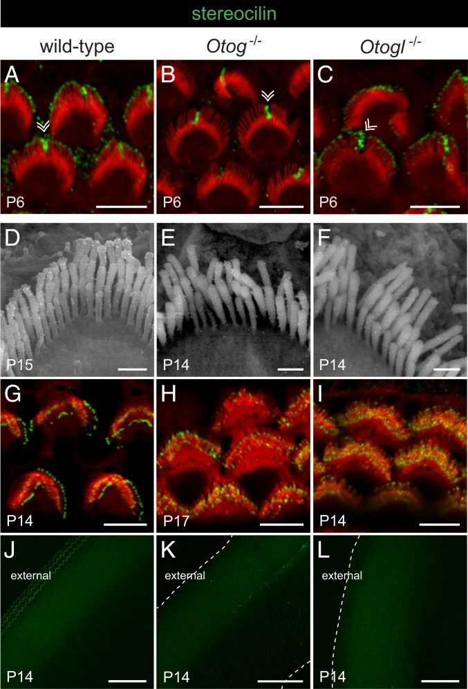
Fig. 7.
Stereocilin in the OHCs and TM of wild-type, Otog−/−, and Otogl−/− mice before and after the onset of hearing. Confocal images (A–C and G–L) and scanning electron micrographs (D–F) of the OHC hair bundles (A–I) and TM (J–L) immunolabeled for stereocilin (gold particles on scanning electron micrographs and green staining on confocal images) and stained for actin (red) on confocal images. Double arrowheads in A–C indicate the transient kinocilium still present in some hair cells on P6. In the P14 wild-type mouse, but not in Otog−/− or Otogl−/− mice of the same age, 3 parallel rows of stereocilin-immunoreactive V-shaped imprints are observed at the anchoring points of the OHC tall stereocilia, on the external side of the TM lower surface (J–L); the dashed lines indicate the edges of the TM in mutant mice. (Scale bars: 5 µm in A–C and G–I, 0.5 µm in D–F, and 50 µm in J–L.)

