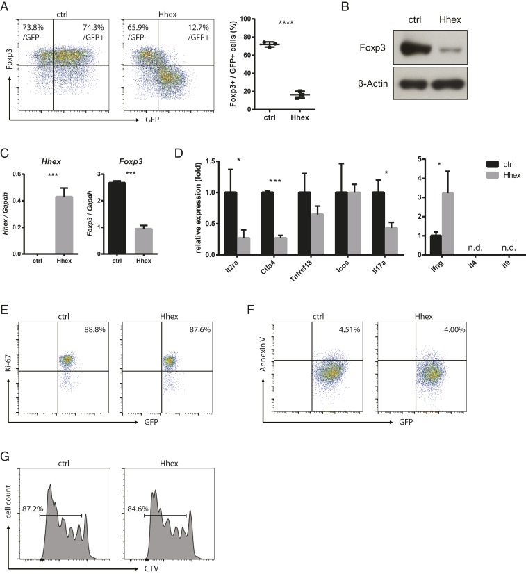
Fig. 2.
Hhex overexpression inhibits the expression of Foxp3 and Treg signature genes. (A–F) Naïve CD4 T cells were stimulated for 24 h and transduced with a retroviral vector expressing GFP with or without (ctrl) Hhex. Cells were cultured for an additional 2 d under iTreg differentiation conditions. Foxp3 expression was detected by flow cytometry, and the ratio of Foxp3+ cells among GFP+ (transduced) cells is shown (A). GFP+ cells were sorted, and the relative amount of Foxp3 protein was detected by immunoblot analysis (B). The expression of Hhex, Foxp3, and other Treg signature genes was measured by qRT-PCR (C and D). Proliferation of the GFP+ cells was determined by Ki-67 staining (E). Apoptosis was measured by Annexin V staining (F). (G) Transduced iTreg cells were stained with CTV and cultured for an additional 4 d in the presence of anti-CD3/CD28 beads and IL-2. All error bars represent the SD, and P values were calculated using Student’s t tests. *P < 0.05, ***P < 0.001, ****P < 0.0001. n.d., not detected.

