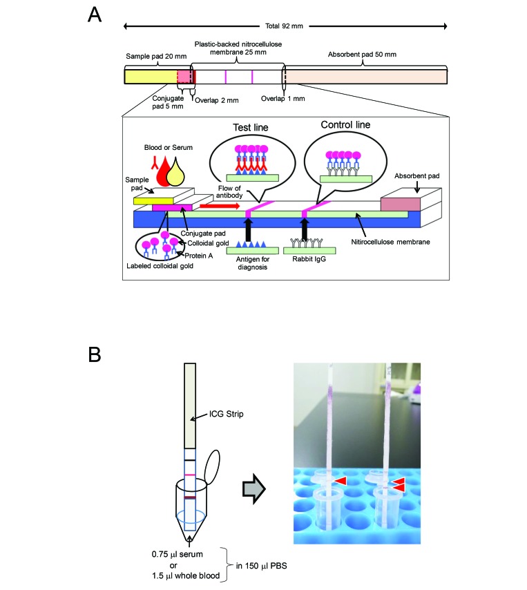
Figure 1.
Detection method and scheme of the immunochromatographic test (ICA) strip. (A) Structure of the ICA strip. Antigens and rabbit IgG were placed at the test lines and control line, respectively. The ICA strip consisted of 4 membrane pads: sample pad, conjugate pad, nitrocellulose membrane, and absorbent pad. The conjugate pad contained the protein A–colloidal gold conjugate. (B) Detection method. Serum (0.75 μL) and whole blood (1.5 μL) were diluted with 150 μL of PBS and then placed in a microtube.

