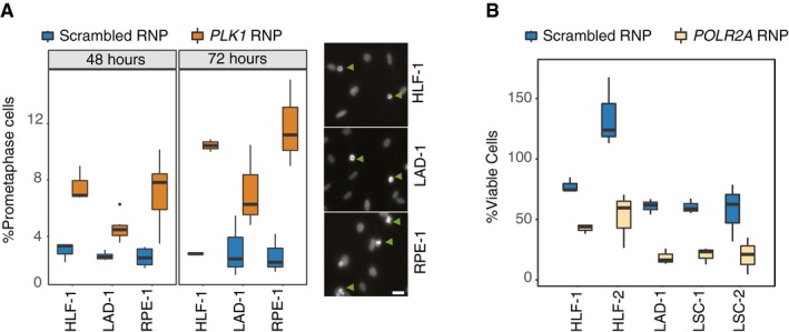
Figure 5. Solid‐phase transfection in primary cells derived from human tumors.

- Solid‐phase transfection of nontargeting (scrambled) or PLK1 targeting RNP complexes into human primary lung fibroblasts (HLF‐1), primary lung adenocarcinoma cells (LAD‐1), or RPE‐1TP53−/− cells. 72 hours post‐transfection, cells were stained with Hoechst and imaged. Boxplots represent values from three independent experiments containing three technical replicates. For all cell lines, P values (scrambled versus Plk1) < 0.05. In the right panels, representative images for each cell line are shown after RNP transfection targeting PLK1. Arrowheads indicate the cells that are arrested in prometaphase due to Plk1 downregulation. Scale bars indicate 20 μm. In the boxplots, centerlines mark the medians, box limits indicate the 25th and 75th percentiles, and whiskers extend to 5th and 95th percentiles. Mann–Whitney U test.
- Two human primary lung fibroblasts (HLF) and three primary tumor cell lines derived from patients suffering from either lung squamous carcinoma (LSC) or lung adenocarcinoma (LAD) were transfected with RNP complexes with scrambled or POLR2A targeting gRNA. Five days post‐transfection, cell viability in each well was measured. Results are from at least three independent experiments containing three technical replicates. In the boxplots, centerlines mark the medians, box limits indicate the 25th and 75th percentiles, and whiskers extend to 5th and 95th percentiles. For all cell lines, P values (scrambled versus POLR2A) < 0.005. Mann–Whitney U test.
