Figure 1.
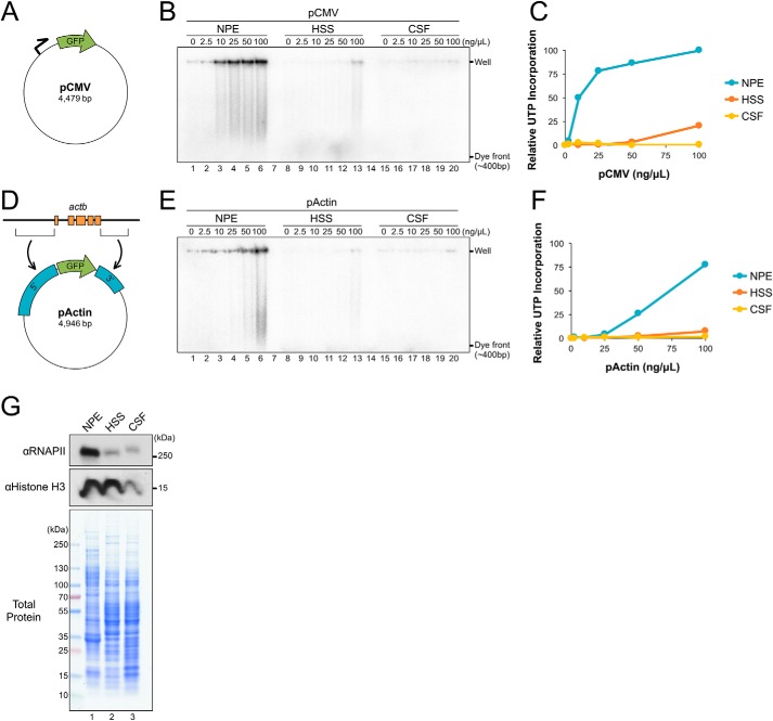
NPE supports robust transcription of plasmid substrates. A, pCMV schematic. Relative location of promoter and GFP are indicated. B, different concentrations of pCMV were incubated in NPE, HSS, or CSF extract in the presence of [α-32P]UTP. Samples were withdrawn at 180 min, resolved by agarose gel electrophoresis, and visualized by autoradiography. C, total UTP incorporation from (B) was quantified and graphed. D, pActin schematic showing the 5′ and 3′ regions cloned from Xenopus actb. E, pActin was incubated in NPE, HSS, or CSF and UTP incorporation was analyzed in parallel to (B) to allow a direct comparison. F, total UTP incorporation from (E) was quantified and graphed relative to peak intensity in (B). G, total protein from each extract was resolved by SDS-PAGE and visualized with Coomassie Brilliant Blue stain or by Western blotting using the indicated antibodies.

