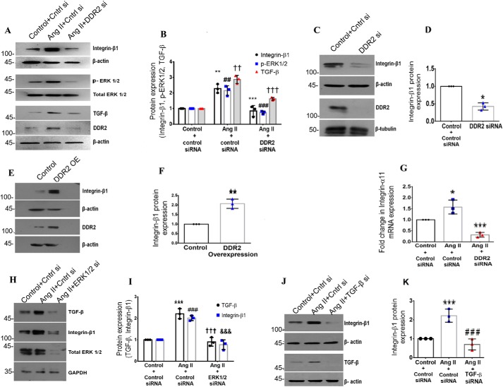
Figure 1.
DDR2 mediates integrin-β1 expression in cardiac fibroblasts exposed to Ang II. A and B, cardiac fibroblasts were transiently transfected with DDR2 siRNA or scrambled siRNA (control). Following exposure of the transfected cells to Ang II for 12 h, integrin-β1 protein expression was examined by Western blot analysis, with β-actin as loading control. **, p < 0.01 versus control; ***, p < 0.001 versus Ang II. Phospho-ERK1/2 activation was examined by Western blot analysis and normalized to total ERK1/2 levels. ##, p < 0.01 versus control; ###, p < 0.001 versus Ang II. TGF-β1 protein expression was examined by Western blot analysis, with β-actin as loading control. ††, p < 0.01 versus control; †††, p < 0.001 versus Ang II. C and D, cardiac fibroblasts were transiently transfected with DDR2 siRNA or scrambled siRNA (control). Following revival and serum deprivation of the transfected cells for 24 h, integrin-β1 protein expression at baseline was examined by Western blot analysis and normalized to β-actin. *, p < 0.05 versus control. E and F, subconfluent cultures of cardiac fibroblasts in M199 were transiently transfected with DDR2 overexpression vector or empty plasmid vector (control). Following revival and serum deprivation of the transfected cells for 24 h, integrin-β1 protein expression was examined by Western blot analysis and normalized to β-actin. **, p < 0.01 versus control. G, cardiac fibroblasts were transiently transfected with DDR2 siRNA or scrambled siRNA (control). Following exposure of the transfected cells to Ang II for 6 h, integrin-α11 mRNA expression was examined by RT-qPCR analysis. 18S rRNA served as the endogenous control. *, p < 0.05 vs. control and ***, p < 0.001 vs. Ang II. H and I, cardiac fibroblasts were transiently transfected with ERK1/2 siRNA or scrambled siRNA (control). Following exposure of the transfected cells to Ang II for 12 h, TGF-β1 and integrin-β1 protein expression was examined by Western blot analysis with GAPDH as loading control. For TGF-β, ***, p < 0.001 versus control; †††, p < 0.001 versus Ang II. For integrin-β1, ###, p < 0.001 versus control; &&&, p < 0.001 versus Ang II. J and K, cardiac fibroblasts were transiently transfected with TGF-β1 siRNA or scrambled siRNA (control). Following exposure of the transfected cells to Ang II for 12 h, integrin-β1 protein expression was examined by Western blot analysis, with β-actin as loading control. ***, p < 0.001 versus control; ###, p < 0.001 versus Ang II. Data are representative of three independent experiments (n = 3). Error bars, S.D.

