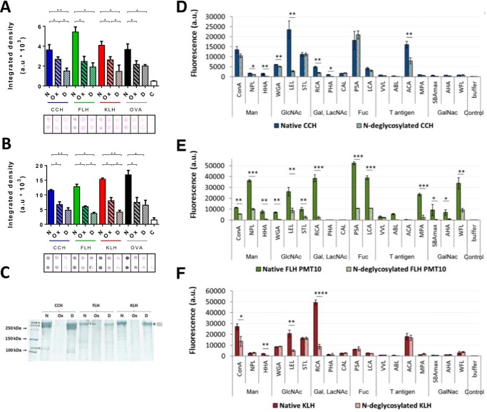
Figure 1.
PNGase F removed partially but significantly N-glycans from hemocyanins. A, dot-blot analysis with PAS-staining method. Native (N), chemically deglycosylated (Ox), and enzymatically N-deglycosylated (D) hemocyanins (CCH, FLH, and KLH) were analyzed. B, dot-blot analysis with lectin staining. 2 μg/ml concanavalin A plus avidin–FAL 1:3000 and NBT-BCIP, which detect N-glycans, were used. A and B, for both analyses ovalbumin (OVA) was used as a deglycosylation control. White bars (C) represent a control of the membranes with all the reagents except hemocyanin. Densitometry quantification of blots was analyzed using ImageJ to quantify integrated density in arbitrary units (a.u). Black borders indicate the cut of dots from the original membrane. Data are shown as the mean ± S.E. Statistical analysis by t test. *, p < 0.05; **, p < 0.01. Representative images are of three independent experiments. C, SDS-PAGE analysis. Native (N), chemically deglycosylated (Ox), and N-deglycosylated (D) hemocyanins were analyzed. Representative gradient acrylamide gel (5–15%) was stained with Coomassie Blue. Molecular mass standards are indicated on the left. In addition, hemocyanin subunits are indicated: CCH (CCH-B, CCHA-1, and CCHA-2), FLH (a single band), and KLH (KLH1 and KLH2). Representative image of three independent experiments. Lectin array blotting of native and N-deglycosylated (D) CCH, (E) FLH, and (F) KLH. Hemocyanins were analyzed by a specific lectin assay for mannose (Man), N-acetylglucosamine (GlcNAc), galactose (Gal), N-acetyllactosamine (LacNAc), fucose (Fuc), T antigen, and N-acetylgalactosamine (GalNAc). Each Alexa Fluor-555–hemocyanin (0.08 mg/ml) was incubated for 1 h at 37 °C with the abovementioned lectins. The binding was quantified in arbitrary fluorescence units (fluorescence a.u.). Data are shown as the mean ± S.E. of one experiment with five replicates. Statistical analysis was done by one-way ANOVA, comparing native hemocyanins versus N-deglycosylated hemocyanins. *, p < 0.05; **, p < 0.01; ***, p < 0.001; ****, p < 0.0001.

