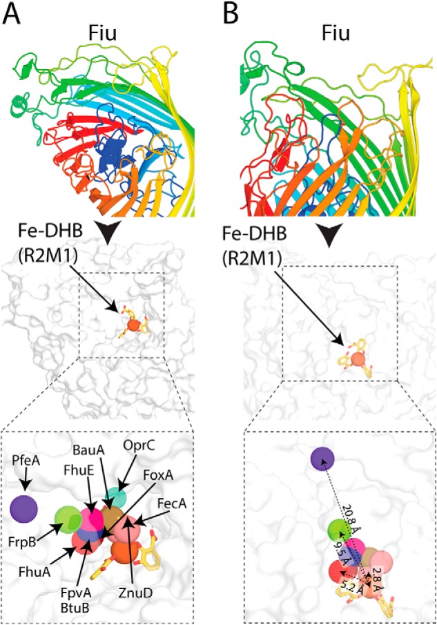
Figure 5.

The location of the Fiu putative external Fe–DHB binding site compared with other TBDT substrate complexes. A, the location of the Fe–DHB complex docked with Fiu compared with that of other substrates bound to in the crystal structures of superimposed TBDTs. Fiu is shown as a cartoon rainbow and in the same view as a white surface representation below. The location of the metal centers of different TBDT substrates are shown as colored spheres and labeled. B, the Fiu Fe-DHB docked complex shown as in A but in a different orientation. Representative distances between Fe–DHB and the TBDT substrate metal ions are shown, colored as in A.
