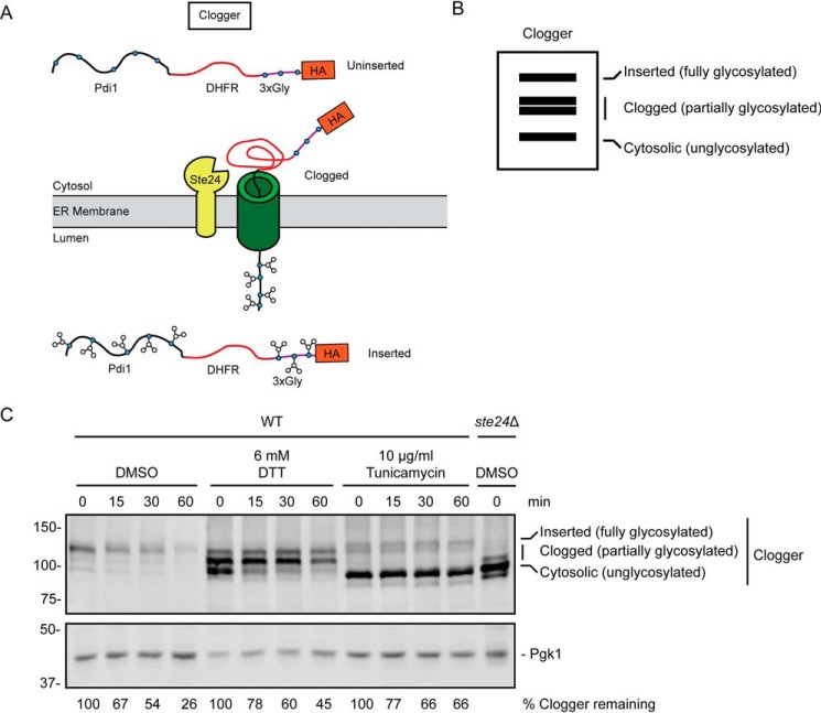
Figure 6.
ER stress impairs degradation of a translocon-clogging substrate of Ste24. A, schematic of Clogger protein prior to (uninserted), during (clogged), and following (inserted) translocon engagement. Clogger consists of Pdi1 (which possesses glycosylation sites), DHFR, three additional glycosylation sites, and an HA epitope. Glycosylated amino acids are depicted as blue circles. B, virtual SDS-PAGE illustrates differential migration of uninserted, clogged, and inserted Clogger. C, cycloheximide chase analysis of yeast of the indicated genotypes expressing Clogger cultured in the presence of 6 mm DTT, 10 μg/ml tunicamycin, or DMSO for 1 h. DTT, tunicamycin, and DMSO were maintained at the same concentration during incubation with cycloheximide. Clogger was detected with anti-HA antibodies. Pgk1 served as a loading control. The percentage of Clogger remaining at each time point (normalized to Pgk1) is indicated below the image. The experiment depicted was performed three times.

