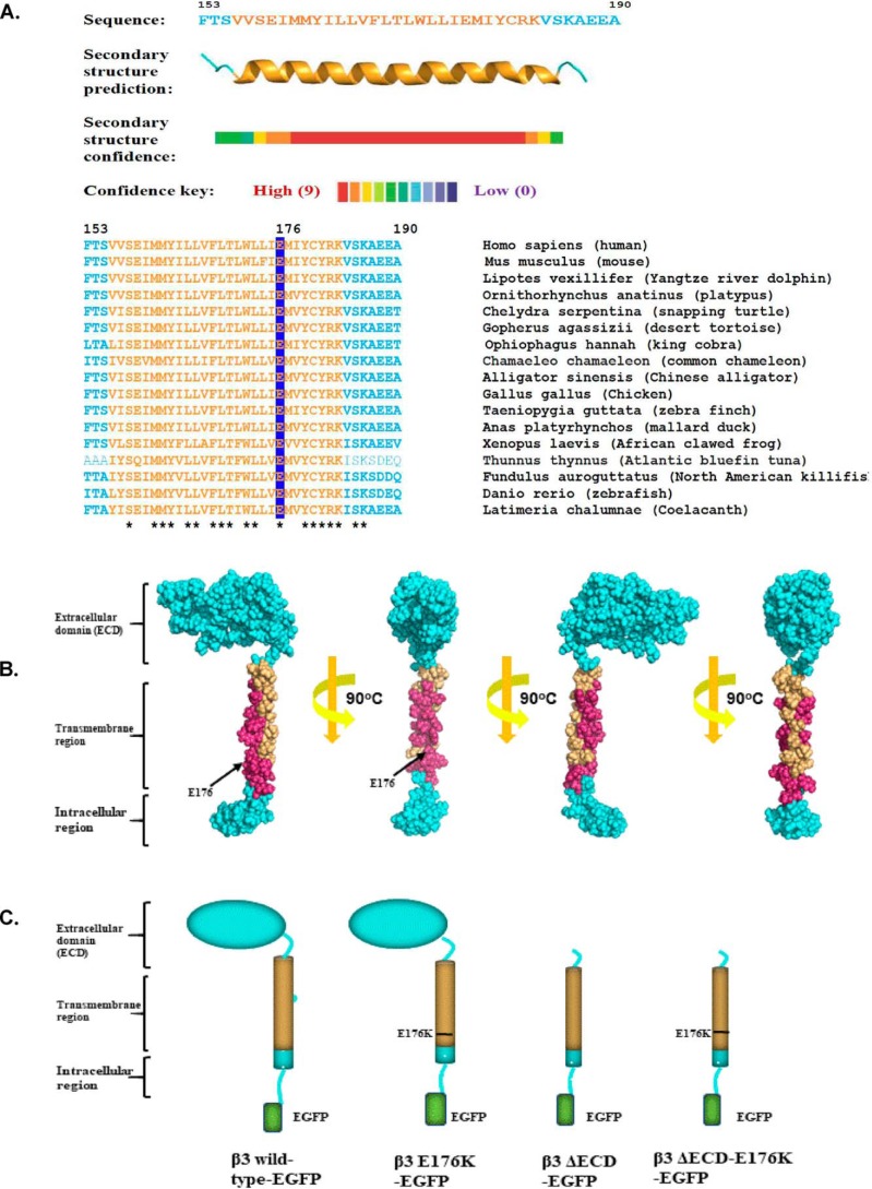
Figure 1.
Sequence analysis of β3-structures. A, secondary structure prediction of the β3-subunit transmembrane region and its sequence alignment among a wide range of vertebrate species. Residue numbers refer to the human sequence. The predicted transmembrane region is colored orange, and the Glu-176 residue is highlighted. Residues fully conserved between species are indicated with asterisks. B, space-filling model of the β3-subunit with the fully conserved residues of the transmembrane region shown in magenta. Note that the helix face containing the Glu-176 residue is fully conserved. Analysis and modeling were as described under “Experimental procedures.” C, cartoon summary of the WT and mutant β3-subunit constructs used in this work and referred to in the text.

