Fig. 1.
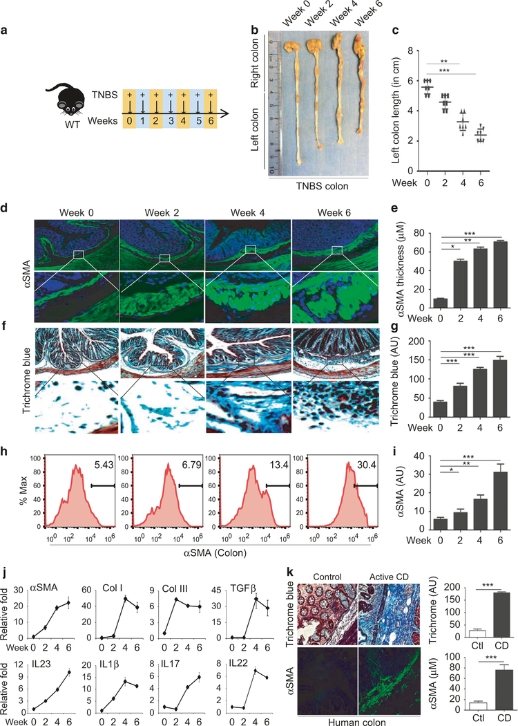
Serial TNBS rectal administration promotes the development of intestinal fibrosis. a Schematic diagram showing the regimen of weekly TNBS treatment given to wild-type mice; b Representative images of colon harvested on week 0, 2, 4, and 6 post-TNSB treatment; c Average length of distal colon, called left colon, plotted in the graph (n= 4–6 mice per group); histological analysis of colon fibrosis shown by representative images of myofibroblasts stained with anti-αSMA antibody (d) and collagen stained by Trichrome blue (f); e Quantification of the thickness of the submucosal αSMA-positive layer and average intensity of Trichrome blue staining at week 0, 2, 4, and 6 (g); h FACS analysis of αSMA-positive cells in the colon, and quantification of αSMA-positive cells (i); qPCR analysis of fibrosis markers and cytokines (j); Data are presented as mean ± SEM (t-test). n= 3–6/group. k Histological analysis of myofibroblast proliferation with αSMA staining and trichrome blue staining for collagen deposition in colon tissue resections from Crohn’s disease patients and controls. Quantification of the intensity of trichrome blue and submucosal αSMA staining in three pairs of surgical specimens. N= 3 *p < 0.05, **p < 0.01, ***p < 0.001

