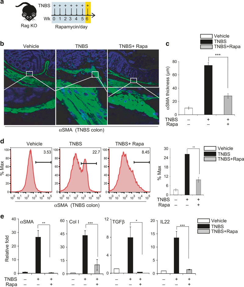
Fig. 5.
Rapamycin attenuates the TNBS-induced fibrotic response in RAG−/− knockout mice, independent of T and B cells. a Diagram showing weekly rectal administration of TNBS and intraperitoneal injection of rapamycin daily (on weekdays) in RAG−/− knockout mice (n= 4–7 mice per group); b Representative images of αSMA staining; c Quantification of the thickness of the submucosal αSMA-positive layer; d FACS analysis of αSMA-positive cells in the colon and quantification of αSMA-positive cells; e qPCR analysis of fibrosis markers and cytokines in purified CX3Cr1+ mononuclear cells from mouse colons. Data are presented as mean ± SEM. n= 3, *p < 0.05, **p < 0.01, ***p < 0.001

