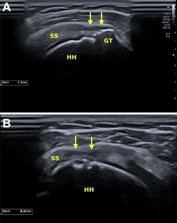
Figure 2.

Ultrasound with a high-frequency (18 MHz) linear transducer: (A) longitudinal and (B) transverse planes of the supraspinatus tendon. Partial-thickness retear of the bursal fibers (yellow arrows), compromising 50% of the tendon. GT, greater tuberosity; HH, humeral head; SS, supraspinatus tendon.
