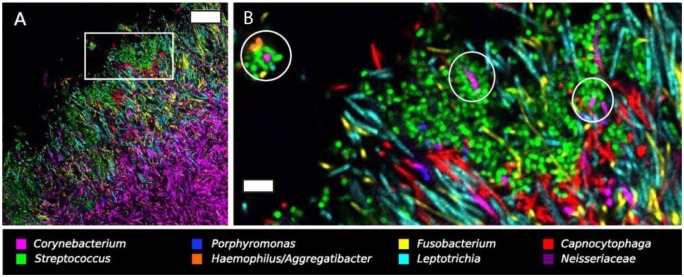
Figure 3.
Systems imaging reveals long-range structure and corncob arrangements in supragingival plaque. (A) Hedgehog structure in supragingival plaque extracted from a healthy volunteer, labeled with fluorescent in situ hybridization probes for 8 genera. Scale bar = 20 µm. (B) Higher magnification view of the region of interest highlighted in A. Circles indicate variously comprised corncob arrangements in hedgehog structures, with coccoid cells (Streptococcus, Haemophilus/Aggregatibacter, and Porphyromonas) arranged around a central filamentous Corynebacterium cell. Scale bar = 5 µm. Reprinted with permission from Mark Welch et al. (2016).

