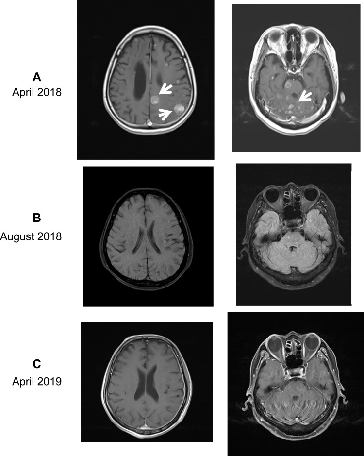
Figure 2.
Post-contrast brain MRI of the head before and after hydrogen inhalation. (A) In April 2018, the patient was found to have multiple metastases (white arrows) in the brain (left, 2.5×3.0 cm and 2.4×2.8 cm for two bigger tumors) and cerebellum (right, 1.3×1.8 cm and 1.2×0.7 cm for two bigger tumors). Also, the third ventricle and lateral ventricles were expanded and had accumulated hydrocephalus. (B) After 4 months of hydrogen inhalation, all tumors visible to the naked eye had disappeared, and the shape of ventricles had returned to normal. (C) After 1 year of inhalation (April 2019), recurrence had not occurred.

