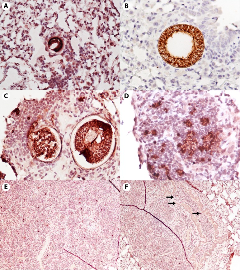
FIG 2.
Histopathology of early murine lung infection. The panels span 72 to 144 h postinfection with C. posadasii strain Silveira. Tissues were immunohistochemically stained with a polyclonal goat antibody against the Coccidioides cell wall antigen, Ag2/PRA, with hematoxylin counterstain. Note that spherules and endospores appear dark red to brown. (A and B) Unruptured first-generation spherules with very few surrounding immune cells at approximately 72 h (A) and approximately 96 h (B). Virtually all are macrophages. (Magnification, ×200 [A] and ×400 [B].) (C and D) There is an enormous influx of PMNs, and also macrophages, once the endospores are released (approximately 120 h). Note the lack of inflammatory cells around the unruptured spherule in panel C. (Magnification, ×200 [C] and ×400 [D].) (E and F) Ongoing recruitment of inflammatory cells to the area and dispersal of endospores, now enlarging back into early spherules (approximately 144 h). In panel F, the top two arrows point to a damaged airway filled with inflammatory cells; the third arrow points to endospores within the damaged airway. (Magnification, ×40 [E and F].)

