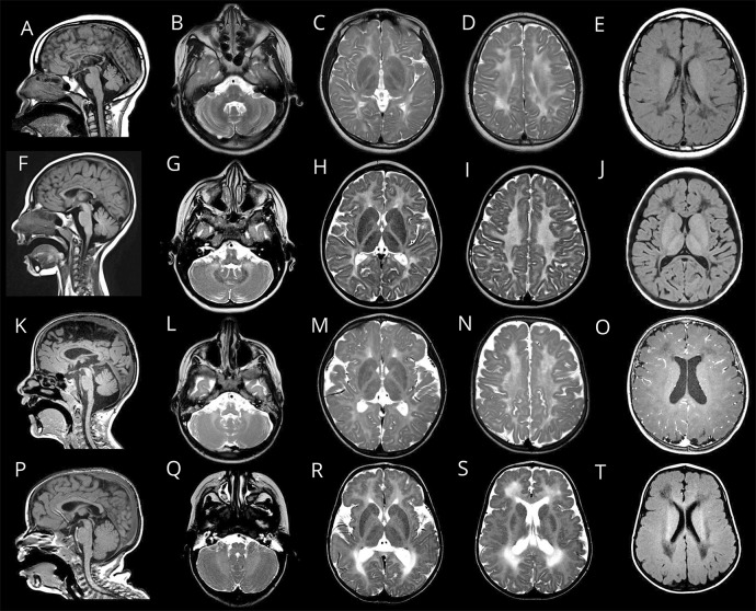
Figure 2. Brain MRI characteristics of 4 patients with POLR3-HLD caused by biallelic POLR1C pathogenic variants.
Sagittal T1 (A, F, K, and P), axial T2 (B–D, G–I, L–N, and Q–S) and axial T1 (E, J, O, and T) images. (A–E) MRI of patient 18 obtained at age 11 years showing diffuse hypomyelination with superimposed areas of pronounced T2 hyperintensity (C and D) and corresponding T1 hypointensity (E). Thinning of the corpus callosum and mild superior vermis atrophy are also seen (A), as well as preserved myelination of the dentate nucleus (B), globus pallidus, anterolateral nucleus of the thalamus, and optic radiation (C). (F–J) MRI of patient 4 obtained at age 5 years showing diffuse hypomyelination with preservation of the dentate nucleus (G), anterolateral nucleus of the thalamus, and optic radiation (H). There is also thinning of the corpus callosum and mild vermis atrophy (F). Areas of marked T2 hyperintensity of the white matter are seen (H and I), with corresponding pronounced T1 hypointensity (J). (K–O), MRI of patient 1 obtained at age 5 years showing a thin corpus callosum (K), relative preservation of myelination of the dentate nucleus (L), and absent T2 hypointensity of the corticospinal tracts in the posterior limb of the internal capsule (M). (P–T), MRI of patient 20.1 obtained at age 3 years showing areas of prominent T2 hyperintensity of the white matter (R and S) with corresponding T1 hypointensity (T), especially in the deep white matter. There is also bilateral frontal polymicrogyria (R, S, and T). POLR3-HLD = RNA polymerase III-related leukodystrophy.

