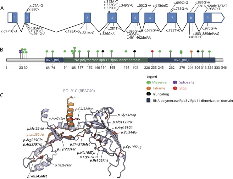
Figure 3. Pathogenic variants identified in POLR1C associated with POLR3-HLD.
(A–B) All reported pathogenic variants and their positions within the POLR1C gDNA (A), with missense variants represented in green, in frame in orange, truncating in black, splice site in purple, and stop in red (B). (C) Missense variants displayed on the structure of the yeast ortholog of POLR1C (RPAC40). Variants previously identified in POLR3-HLD are represented in italic, whereas newly identified variants are shown in bold. The p.Lys295del is shown in orange. The p.Thr26Ile, p.Thr27Ala, and p.Pro30Ser variants have not been represented because they are not visible in the crystal structure of RPAC40 (PDB 5M5W).19,20,38–40 POLR3-HLD = RNA polymerase III-related leukodystrophy.

