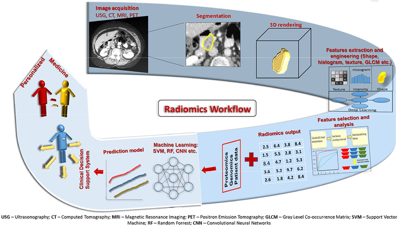
Fig. 2.

The workflow of Radiomics. The first step in the process is selection of the region of interest through manual or (semi)automated segmentation on acquired/archived standard of care radiographic images. Selecting the region of interest on these series of images forms a 3D rendering of the lesion. Subsequently, radiomics features are extracted from this region of interest. In the next step, statistical analysis is used to determine the most pertinent features from all the extracted features. These judiciously chosen features are then combined with the patient’s clinical data to build a prediction model with the help of machine learning tools. Furthermore, this model is validated against an unknown data set to prove its accuracy. Consequently, the radiomics model can form a part of the clinical decision support system to personalize medical care for the patients in the future.
