
Figure 1. Flair and DWI imaging.
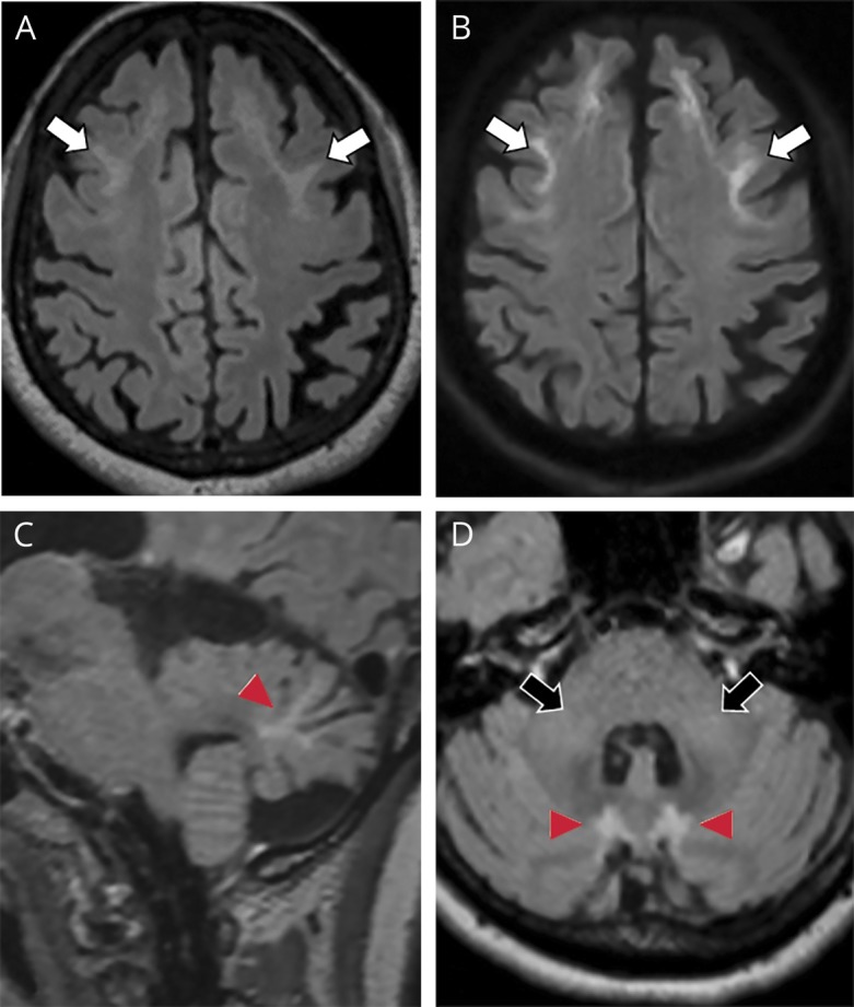
(A) Axial fluid-attenuated inversion recovery (FLAIR) and (B) diffusion-weighted imaging (DWI) of the supratentorial brain show high FLAIR signal with corresponding high DWI signals involving the corticomedullary junction of bilateral frontal lobes (white arrows, A and B). (C) Sagittal FLAIR and (D) axial FLAIR of the posterior fossa show FLAIR hyperintensities within paravermal regions (arrowheads, C and D) and in bilateral cerebellar peduncles (black arrows, D).