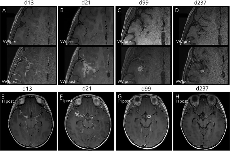
Figure 2. Serial vessel wall MR and T1 SPACE images.
Pre- and postcontrast VWI images of the right frontotemporal sulci at the level of the right sylvian fissure are shown at (A) 13, (B) 21, (C) 99, and (D) 237 days from initial presentation. (A) At 13 days, leptomeningeal and vessel wall enhancement is present. (B) At 21 days, parenchymal enhancement consistent with cerebritis and evolving infarctions centered around the infected vessels were present. (C) At 99 days, an enhancing tuberculoma in the right sylvian fissure developed. Concurrently, there was resolution of the parenchymal enhancement and decreased vessel wall enhancement. (D) By 237 days, there was complete resolution of the vessel wall enhancement leaving only residual wall thickening and a contracting tuberculoma. (E–F) At days 13 and 21, imaging shows thick enhancing basilar exudates in the sylvian fissures bilaterally. (G–H) By day 99, a ring-enhancing tuberculoma in the left suprasellar cistern developed, which gradually contracted by day 237. VWI = vessel wall MR imaging.

